Bringing the resolution revolution to Groningen
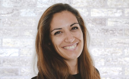
When Cristina Paulino started working on cryo-electron microscopy almost ten years ago, no one could have foreseen it would become one of the hottest fields in science. The field was even the subject of this year’s Nobel Prize for Chemistry. Paulino has brought this new technique to the University of Groningen.
She has just published a paper in the prestigious journal Nature, on her work in her previous position at the University of Zürich. ‘Over the last three years, more than two hundred papers based on cryo-electron microscopy have been published in top journals like Nature, Science or Cell.’ It’s the result of the resolution revolution, a rapid advance in electron microscopy technology and software that allows scientists to resolve the structure of complex proteins at the atomic level.

All you need to join in is a EUR 3-6 million microscope, some EUR 200,000 per year for the maintenance contract, another few 100,000 for a powerful image processing cluster and some very experienced operators. ‘Getting the right staff to operate such a machine is a big problem at the moment’, says Paulino. She currently spends a lot of time at the microscope. ‘You need experience to do it well’, she explains.
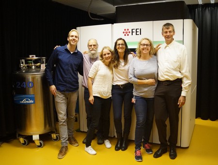
Four years ago, only a few big national labs, like those funded by the US National Institutes of Health or the German Max Planck Institutes, could afford a good cryo-electron microscope. But as these become more user-friendly and affordable, more labs are getting them – and need qualified staff to operate them. In the Netherlands, two of the best cryo-electron microscopes for life sciences can be found at the Netherlands Centre for Electron Nanoscopy (NeCEN) in Leiden. Other high-end microscopes have recently been installed in Utrecht, Delft and Maastricht. With the recruitment of Paulino, there is now one in Groningen too, which has been up and running since June, and is producing its first results.
Shock frozen
As Principal Investigator, Paulino is responsible for the machine. But she emphasizes she’s not only a microscopist: ‘I’m a biochemist with an interest in membrane proteins, especially their structural biology. So I want to know what they look like.’ But she’s trained in all biochemical techniques. In 2008, she started her PhD project in the lab of Werner Kühlbrandt, one of the pioneers of cryo-electron microscopy. ‘At that time, no one expected the technique to become so big.’

To achieve atomic resolution, every step in the process must be completed correctly. And there are lots of steps. First, the sample is shock frozen by dipping it in liquid ethane cooled with liquid nitrogen (some 180 degrees below zero). ‘The protein you want to study is mounted on a grid in a thin film of water. The freezing process should be very quick, so that no destructive ice crystals can form.’
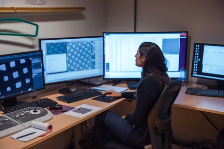
The vitrified sample is then placed in the microscope. ‘You need steady hands for this – otherwise you can damage the machine. In some labs, only two or three specialists are allowed to do this.’ The microscope is then aligned and programmed to scan the grid in the right pattern. This can take the best part of a day, but from then on the data acquisition is fully automatic. ‘This is a recent breakthrough. The software to run the machine has become much more user-friendly.’
Experience
But during these preparations, all parts of the microscope have to be aligned to perfection. This is a job you can’t learn in a three-week course. ‘You need years of experience, and will have encountered lots of problems.’ And as already mentioned, experienced operators are scarce. Currently, Paulino is assisted by Gert Oostergetel. ‘He retired two years ago, but is staying on to help me out. He has a tremendous amount of experience with electron microscopy.’

The machine scans from a few thousand to a million proteins on the grid. The next step is to add together the images from all proteins, which gives you the full structure. ‘On a good day, you can record up to four terabytes of data. Only astronomers have more data to process, store and analyze.’ Paulino has already spent over EUR 100,000 on computational power. ’And I need more. I’ve become quite the computer buff over the last few years.’
Privilege
Paulino currently spends quite a bit of her time working at the microscope. ‘But being a PI for the first time is also time-consuming.’ So she hopes to find a good operator soon. Not that she’s complaining about her current position: ‘Having access to a machine of this quality, which can go to a resolution of 3 Angstrom, is quite a privilege. For most scientists, getting time on a microscope is the major problem. I have all the access I want.’

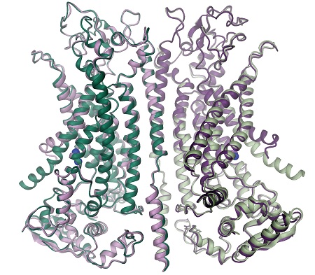
On 13 December, she published a paper in Nature on her work in Zürich. It describes the structure of a protein that looks like a scramblase, a protein that sits in the cell membrane and is able to flip phospholipids (the building blocks of the membrane) between the inner and outer layer. She discovered it works like an ion channel, with two pores that open in the presence of calcium.
Competition
‘So the question was, How can a scramblase function as a channel? Our high-resolution structure enabled us to answer that.’ A calcium binding site in a cavity of the transmembrane portion of the protein complex can induce a conformational change which opens the pore.
This shows the kind of results which can be obtained with the atomic resolution of proteins. Since starting work in Groningen, Paulino has already worked with colleagues at the Linnaeusborg and in Germany and Switzerland to resolve the structure of other proteins. ‘I’m afraid I can’t provide much detail on what I’m doing, because the competition is immense.’ We predict some great papers next year.
See also:
In search of the basis of life
Nobel Prize for Chemistry 2017
Reference:
Cristina Paulino, Valeria Kalienkova, Andy K. M. Lam, Yvonne Neldner & Raimund Dutzler: Activation mechanism of the calcium-activated chloride channel TMEM16A revealed by cryo-EM. Nature, 13 December, DOI 10.1038/nature24652
| Last modified: | 02 May 2019 09.12 a.m. |
More news
-
10 June 2024
Swarming around a skyscraper
Every two weeks, UG Makers puts the spotlight on a researcher who has created something tangible, ranging from homemade measuring equipment for academic research to small or larger products that can change our daily lives. That is how UG...
-
21 May 2024
Results of 2024 University elections
The votes have been counted and the results of the University elections are in!

